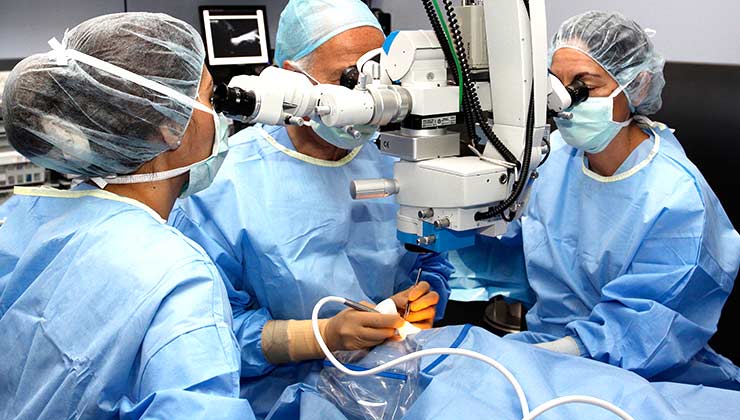
Retinal detachment can only be fixed through surgery and unfortunately, not many people really understand how retinal detachment surgery is done or what it entails. In fact, there are multiple ways of performing retinal detachment surgery depending on which is best for the patient’s diagnosis. For a more in-depth look at the procedure visit https://asiaretina.com/retinal-detachment/.
Meanwhile, here are a few things you should probably know about before deciding to get retinal detachment surgery:
1. The retina is one of the most important parts of the eye.
The retina is the inner layer located at the back of one’s eye and is one of the most important parts of the eye, as it is responsible for capturing the light passing through your eye and then sending that signal to the brain for it to make sense of. Without the retina, a person is rendered blind as the light signals do not get sent to the brain.
2. Retinal detachment is a serious eye condition and needs treatment ASAP.
Since the retina is an essential part of your eye, if it ever gets detached, then there is a possibility that a patient may lose his/her vision on that eye permanently. To avoid this from happening, it’s important that the patient is aware of the symptoms of retinal detachment and to seek help as soon as possible if he/she exhibits one or more of these symptoms.
3. There isn’t any pain once your retina starts getting detached.
However, you will start to notice your vision becoming blurry or cloudy, as well as suddenly seeing lots of floaters, which are small spots or lines that drift into your field of view. You will also experience some photopsia, in which there are sudden flashes of lights that show up in one or both eyes. Some people also experience their vision to have a dark “curtain” over it, either on the sides (peripheral) or even in the middle of their field of view.
4. A retinal tear can happen before detachment.
Before the retina starts to detach, it is quite common to experience a retinal tear. This describes a phenomenon in which small cuts appear on the retina, causing floaters to appear in the field of vision. It has very similar symptoms to retinal detachment, so if one experiences these symptoms, it is important that you see an eye doctor right away.
5. There are three possible reasons for the retina to detach.
The first and most common reason is when there is a retinal tear, which in turn lets fluid pass through and gather under the retina. The weight of the fluid pulls the retina away from the eye tissues holding it together, causing the retina to detach. This is usually caused by the vitreous, the gel-like material that fills the eye, starting to change into a liquid. This is what’s called a rhegmatogenous retinal detachment.
Another reason is tractional retinal detachment. This is when there is the presence of scar tissue around your retina, which can then start to pull on it, causing it to detach. These scar tissues usually appear due to diabetes, which can damage the blood vessels on your retina.
The last possible reason is an exudative retinal detachment, which is when fluid gathers behind the retina but without the presence of retinal tears. This usually happens due to aging, an injury or inflammation.
6. Some people are more at risk for retinal detachment than others.
If a person has any of the following:
- Is over the age of 50
- A family member who had retinal detachment
- Extreme myopia (nearsightedness)
- An eye injury
- Undergone cataract removal surgery or other eye surgeries
- Diabetic retinopathy
then he/she has a higher risk of retinal detachment in the future. You should see an ophthalmologist for an eye checkup if you have any of the above risk factors.
7. Retinal detachment can be diagnosed by your eye doctor.
The process usually involves the doctor applying eye drops to dilate your pupil. He/she will then examine the back of your eye using a special instrument that takes clear and detailed pictures of your retina.
Another way to diagnose retinal detachment is using ultrasound imaging. This is done if some bleeding has occurred, and it’s become harder to use the previous method. Even if you only experience symptoms in one eye, the doctor will check both of your eyes for retinal detachment to be sure.
8. Retinal detachment needs surgery to be repaired.
Before retinal detachment surgery is done, a general or local anaesthetic is first injected around the patient’s affected eye so that there is very little pain or discomfort during the procedure. A retinal detachment surgery operation usually takes around 1.5 hours to finish.
Depending on the type of retinal detachment, the eye doctor can utilise different methods to repair a retinal detachment.
For any retinal tears or holes, the surgeon can use either laser surgery or a freezing technique (cryopexy) to repair it. In laser surgery, the surgeon will use a laser beam to burn around the tear, effectively welding the tear closed. The surgeon may also freeze the retinal tear together to achieve a similar result.
For the retinal detachment itself, the surgeon can utilise three different techniques. The first one, called pneumatic retinopexy, involves the injection of an air bubble into the center of the eye. This bubble effectively pushes the retina against the wall of the eye, stopping fluid from building up behind it.
Another technique, called vitrectomy, involves the removal of the vitreous and any other tissue that’s pulling on the retina. A substance is then injected to help flatten the retinal wall.
The last technique, known as scleral buckling, is done by using a piece of silicone and sewing it to the sclera. This reduces the force applied by the vitreous and relieves that area from detachment.
9. It can take several months to recover from retinal detachment surgery.
Before you decide to take the surgery, do not be afraid to ask your doctor and surgeon about the possible risks and benefits of the procedure. Although complications are possible, and you may need to undergo a follow-up surgery in the future, the benefit of potentially repairing your vision cannot be understated. The usual recovery period can take 3 to 6 months, but many patients are satisfied with the results so it’s up to you to decide if the benefits outweigh the risks involved.
Asia Retina – Eye specialist (Ophthalmologist) in Singapore, Dr Claudine Pang
#15-10 The Paragon, 290 Orchard Rd, 238859
+65 6732 0007
https://asiaretina.com

